
Figure 3.
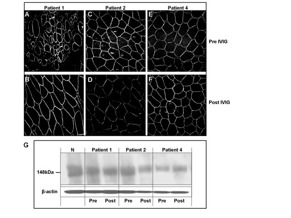
Expression of α-dystroglycan in quadriceps muscle from HIBM patients. A-F. Immunohistochemistry using antibodies to IIH6. No consistent difference in staining was apparent before (A,C,E) compared with after (B,D,F) IVIG treatment. The specimen shown in A was sampled from a deteriorating area. G. Immunoblots of muscle from a normal individual (N) and HIBM patients 1, 2, and 4, labeled with antibodies to α-dystroglycan. No consistent difference in IIH6 staining (reflecting the sialylation status of α-dystroglycan) was apparent before (Pre) compared with after (Post) IVIG treatment. β-Actin bands provide an indication of the level of protein loading.