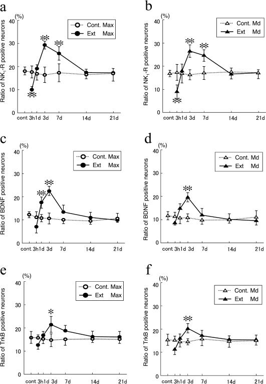
Fig. 3.
The ratio of NK1-R- (a), BDNF- (b), and TrkB-immunoreactive neurons per PGP-9.5-immunoreactive neuron in the maxillary (a, c, e) and mandibular (b, d, f) nerve regions between 3 hr and 21 days after extraction. Cont., control; Ext, extracted groups; Max, maxillary nerve region; Md, mandibular nerve region. Data are expressed as the mean±SD. Control group n=4. Experimental group n=7. ** p<0.01, * p<0.05.

