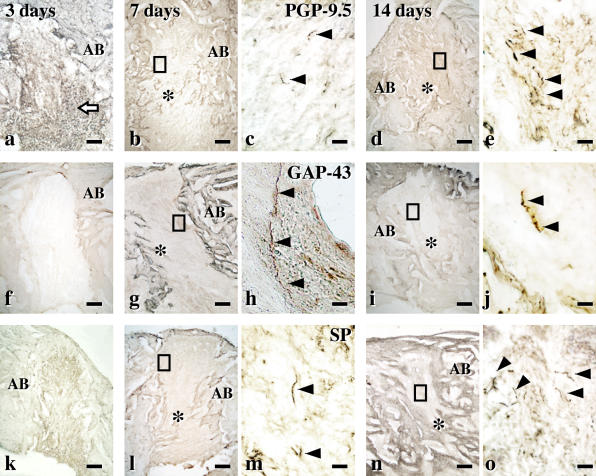
Fig. 4.
Reinnervation during the healing of extracted sockets. Immunohistochemical images for PGP-9.5 (a–e), GAP-43 (f–g) and SP (k–o) at the tooth extracted socket. (a, f, k) Three days after extraction, the extracted root sockets are filled with inflammatory cells (arrow) and (a) no PGP-9.5-immunoreactive nerve fibers are seen. (b, c, g, h, l, m) Seven days after extraction. (b, g, l) The root socket is filled with fibrous tissues. (h) High-power images of the square area in (g). GAP-43-immunoreactive nerve fibers (arrowheads) appeared in the extracted socket. (m) Some SP-immunoreactive nerve fibers (arrowheads) is seen. (d, e, i, j, n, o) Fourteen days after extraction. (d, i, n) The extracted sockets are partly filled with new bone. (o) SP-immunoreactive nerve fibers (arrowheads) was often observed in the extracted root sockets. AB, alveolar bones; *, new bone. Bars=200 µm (a, b, d, f, g, i, k, l, n) and 20 µm (c, e, h, j, m, o).

