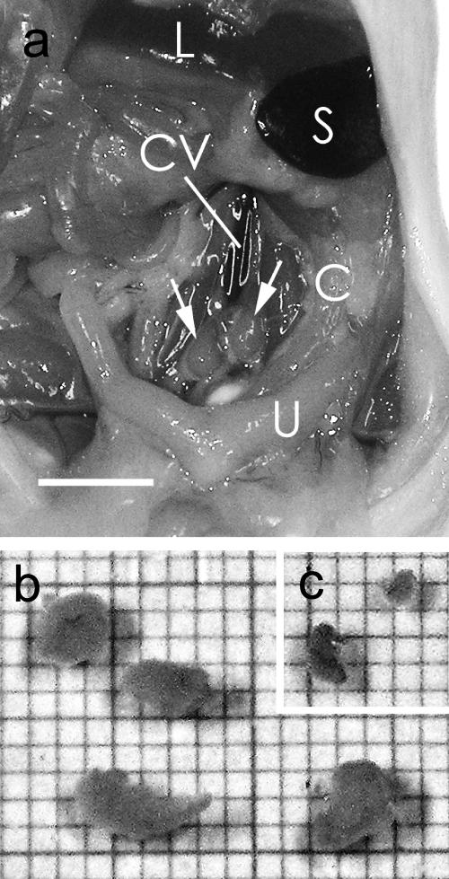
Fig. 2.
a: Enlarged iliac lymph nodes (arrows) from a BALB/c mouse injected with antigen emulsion. C, colon; L, liver; S, Spleen; U, uterus; CV, caudal vena cava. Bar=5 mm. A small portion of the antigen emulsion was found in the sacral lymph node. b: Enlarged iliac lymph nodes in culture medium from two BALB/c mice 14 days after injection of the antigen emulsion. The scale of the graph paper is 1 mm. c: Normal iliac lymph nodes from an age-matched BALB/c mouse.

