Fig. 1.
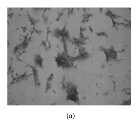
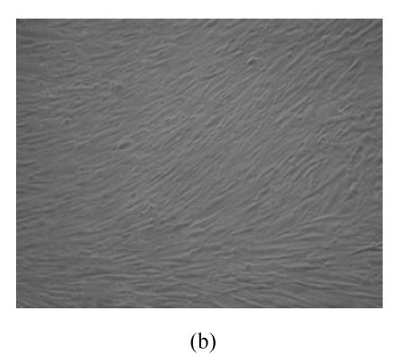
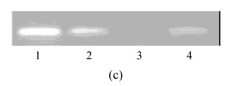
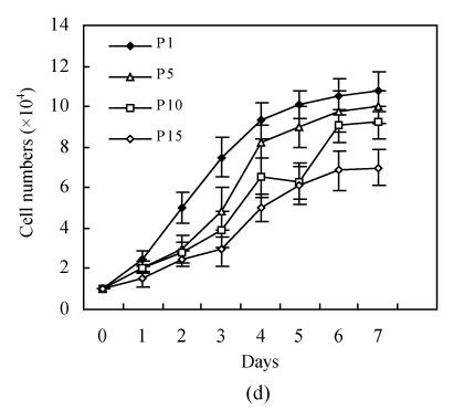
Ex vivo growth morphology of resuscitated hMSCs. (a) Colonies of post-cryopreserved hMSCs stained with Wright-Giemsa (×100); (b) Confluence of post-cryopreserved hMSCs (×100); (c) RT-PCR for Oct4 (line 1 for passage 1 of resuscitated hMSCs, line 2 for passage 5 of resuscitated hMSCs, line 3 for control cells (fibroblast cells) and line 4 for passage 10 of resuscitated hMSCs); (d) The growth curves of hMSCs at passage 1 (P1), 5 (P5), 10 (P10) and 15 (P15) of post-cryopreservation


