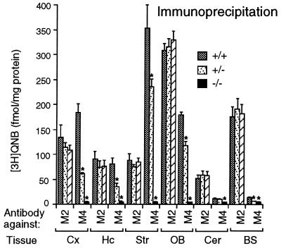
Figure 3.
Immunoprecipitation analysis of muscarinic receptor expression. Immunoprecipitation studies were carried out as described in Materials and Methods, using [3H]QNB-labeled receptors solubilized from the indicated brain regions of wild-type and M4 receptor mutant mice. M2 or M4 muscarinic receptors were immunoprecipitated with subtype-specific anti-M2 or anti-M4 rabbit antisera, respectively. Cx, cerebral cortex; Hc, hippocampus; Str, striatum; OB, olfactory bulb; Cer, cerebellum; BS; brain stem. Data are given as means ± SD (n = 3–4 for each dose and genotype). ∗, P < 0.001 (Student’s t test).

