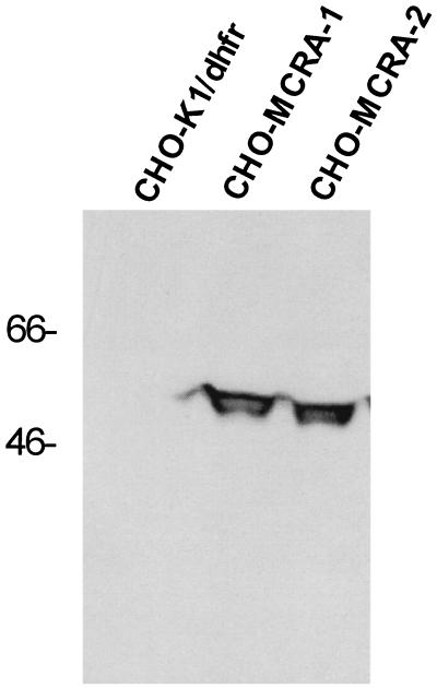
Figure 2.
Western analysis of CHO-K1/dhfr− parental cells and transfected cell clones expressing MCRA. Western analysis was performed with standard protocols as described by Sambrook et al. (17). Proteins were resolved by electrophoresis by SDS/10% PAGE and transferred to nitrocellulose. A rabbit anti-MCRA antibody was used at a 1:4,000 dilution to detect MCRA on nitrocellulose membranes in combination with a horseradish peroxidase-conjugated anti-rabbit antibody and the enhanced chemiluminescent visualization reagent (ECL, Amersham). Molecular weight size markers are indicated in kDa.

