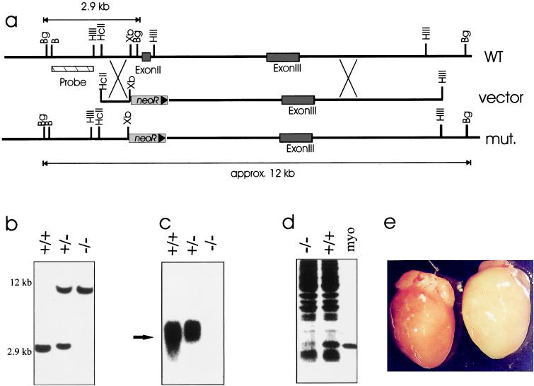
Figure 1.
Targeting strategy and molecular verification of myoglobin disruption. (a) Structures of the WT and mutated alleles (mut) and the targeting vector are shown. (Restriction sites: B, BamHI; Bg, BglII; HIII, HindIII; HcII, HincII; Xb, XbaI. neoR, neomycin resistance gene). (b) Southern blot analysis of BglII-digested DNA from WT (+/+), heterozygous (+/−), and homozygously mutated (−/−) mice. The BamHI-HindII-fragment indicated in a was used as a probe. Hybridizing fragments of 2.9 kb (WT allele) and 12 kb (mutated allele) were detected. (c) Northern blot analysis of cardiac RNA isolated from the three genotypes. (d) SDS/PAGE analysis of protein patterns of myo−/− and WT hearts. Two-hundred micrograms of cardiac proteins were separated on a 12.5% SDS gel and were stained with Coomassie brilliant blue. Five micrograms of horse myoglobin were loaded as control. (e) Morphology of myoglobin-deficient hearts. WT (left) and myo−/− hearts (right) were perfused free of blood and were photographed.

