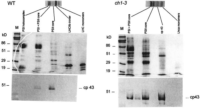
Figure 3.
Second-dimension denaturing gel electrophoreses of wt and ch1-3 thylakoid membranes and immunoblot analysis with anti-CP43. The nondenaturing green-gel is shown at Top. The indicated pigmented bands were electrophoresed on denaturing gels, stained with Coomassie, and are shown in the Middle. Bottom is an immunoblot of the denaturing gels reacted with the anti-CP43 antibody.

