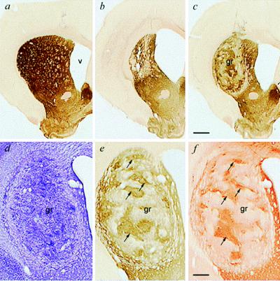
Figure 2.
Photomicrographs of striatal graft survival and integration. (a) Intact neostriatum in a control rat. (b) Unilateral quinolinic acid lesions induce massive neostriatal cell loss, striatal atrophy, and enlargement of the lateral ventricle. (c) Striatal transplant surviving in the lesioned striatum, increasing total striatal volume and reducing ventricular expansion. (a–c) All acetylcholinesterase histochemistry at low magnification. (d–f) Higher-magnification view of the same striatal graft visualized in adjacent sections with cresyl violet to visualize cell bodies (d), acetylcholinesterase to visualize the characteristic cholinergic neuropil of the striatum (e), and DARPP-32, a receptor marker predominantly located on striatal neurons (f). Note the good survival and distinctive patchy internal organization of the grafts, with the striatal-like compartment being aligned in adjacent sections (arrows). [Bars = 1 mm (c) and 250 μm (f).]

