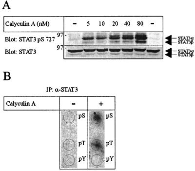
Figure 1.
(A) CA induces serine phosphoryaltion of STAT3 in vivo. Resting, antigen-specific human CD4+ T cells were incubated with increasing concentrations of CA (5–80 nM) for 60 min, lysed in lysis buffer, applied to SDS/PAGE as described in Materials and Methods, and immunoblotted with anti-STAT3 pS727 (Upper), stripped and reblotted with anti-STAT3 (Lower). (B) Amino acid analysis of 32P-orthophosphate-labeled STAT3. 32P-labeled proteins of interest, representing STAT3 proteins from cutaneous T lymphoma cells incubated with or without CA for 60 min, were cut out from the poly(vinylidene difluoride) membrane, hydrolyzed in 6 M HCl, and separated by thin-layer electrophoresis. The 32P-labeled phosphoamino acids were detected by autoradiography as described in Materials and Methods.

