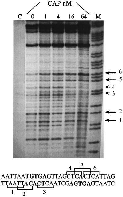
Figure 6.
Sequencing gel of calicheamicin induced cuts on the selected DNA fragment (5′-CATGGCGCAACGCAATTAATGTGAGTTAGCTCACTCATTAGGCACCCTAGGTCTAG-3′) containing the CAP-binding sequence in the presence of increasing amounts of CAP. DNA is labeled 3′ on the bottom strand. [calicheamicin] = 5 μM, [CAP] = 0–64 nM; A + G, purine marker. The large arrows indicate drug-binding sites stimulated by the DNA–protein complex formation; the small arrows indicate inhibited sites. The DNA bases in bold are in direct contact with CAP.

