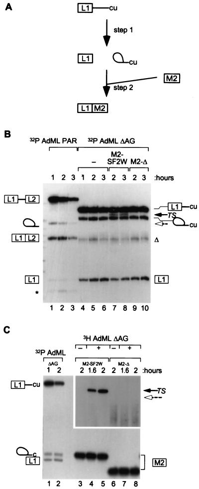
Figure 2.
ESE dependence of bimolecular exon ligation. (A) A scheme of the trans-splicing assay is shown, with the 5′ leader exon denoted by the boxed L1 and the intron by a line ending with “cu” (17). After incubation, to allow the first step of splicing to occur, IgM M2 exon (with a 3′ splice site) was added as a 3′ substrate. (B) AdML ΔAG RNA was 32P-labeled and incubated in nuclear extract for the times shown (lanes 4–6). 3H-labeled 3′ exons were added for the second and third hours of the reactions (lanes 7–10). The trans-spliced mRNA (TS) is shown by a black arrow. The expected position of trans-splicing with M2-Δ is marked by a dashed open arrow. Control splicing in cis between AdML PAR exons L1 and L2 is shown in lanes 1–3. (C) In a reciprocal labeling experiment, the 3′ exons were 32P-labeled, and the 5′ fragment was made with [3H]GTP. Controls with 32P-labeled AdML ΔAG pre-mRNA are shown in lanes 1 and 2. Other controls omitting the 3H-labeled 5′ RNA are shown in lanes 3 and 6. A longer exposure of the trans-spliced products is shown. In B (and in subsequent figures), the asterisk indicates an artifactual band characteristic of the AdML PAR substrate (11, 17); the triangle indicates an artifactual product that appears to derive from the AdML ΔAG lariat intermediate or from use of an alternative branch site.

