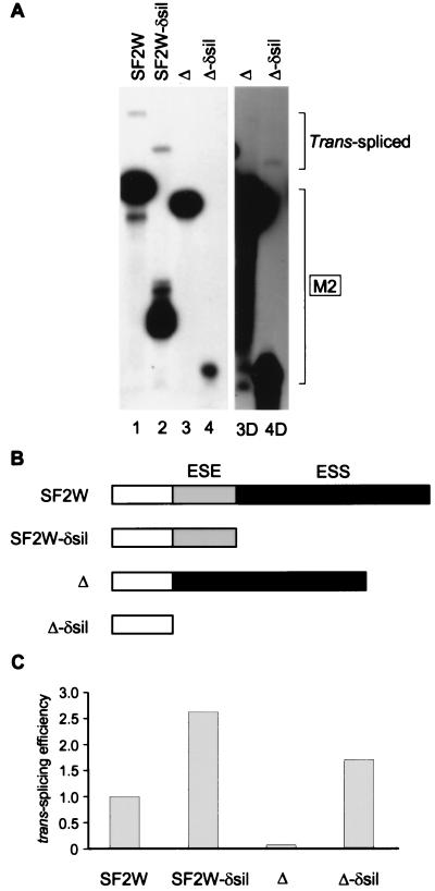
Figure 4.
Interaction between enhancer and silencer elements in trans-splicing. (A) 3H-labeled AdML ΔAG pre-mRNA was incubated for 1 hr in nuclear extract. Then, the indicated 32P-labeled 3′ M2 exons with or without an ESE or a silencer were added. The lower and upper brackets on the right show the positions of the 3′ exons and of the trans-spliced mRNAs, respectively. A longer exposure of lanes 3 and 4 is shown (lanes 3D and 4D). (B) Schematic structure of the different 3′ M2 exons. The ESE is shaded gray, and the silencer segment (ESS) is shaded black. (C) Relative trans-splicing efficiencies of the four 3′ exons. The data from A were quantitated by densitometry and normalized for loading and differential labeling of the 3′ exons. The efficiency of the SF2W 3′ exon was arbitrarily set at 1.

