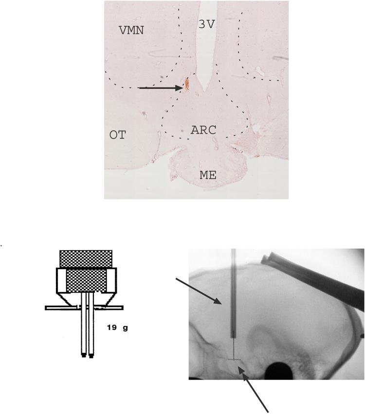
Fig. 4.

Montage of 10x images of the mediobasal hypothalamus of one representative monkey showing the posterior probe placement with DAB staining. The DAB injection site is located near the bottom of the 3rd ventricle of the mediobasal hypothalamus. The anterior site would have been located 2 mm in front of this point. In addition, the design of the headpiece holding the guide cannulae, and a lateral roentgenogram from the same animal (#4), are shown. The xray was obtained at final cannula placement and indicates the cannulae barrels (single-headed arrow). The double-headed arrow indicates the pencil mark on film, which marks the midpoint of the edges of the sella turcica. The single arrowheads indicate the anterior and posterior edges of the sella turcica, which are the critical landmarks for positioning of the guide cannulae. The guide cannulae are positioned at equal distance from the anterior and posterior points. The microdialysis probe extends several millimeters beyond the end of the guide tubes. All of the data reported for the estrogen+progesterone experiments are from animals with similar probe locations. This location contains arcuate nucleus neurons. Abbreviations: ARC-arcuate nucleus; ME- median eminence; OT- optic track; VMN- ventromedial nucleus; 3V- third ventricle.
