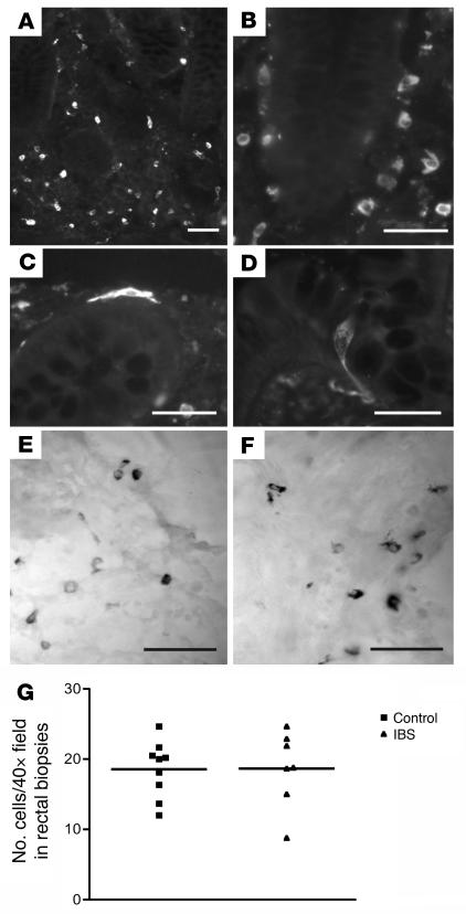
Figure 3. Tryptase immunoreactivity in rectal biopsies from control (A, D, and E) and IBS patients (B, C, and F).
Tryptase was mostly localized to cells found throughout the lamina propria (A–C) and around the base of rectal mucosal crypts (C). Some cells (e.g., C), had obvious processes at either the apical and/or basal poles, and often these cells displayed intense immunoreactivity compared with neighboring cells (images in B and C were taken under identical exposure conditions and reflect differences in intensity). Occasional tryptase-immunoreactive cells were found in the epithelium of both control (D) and IBS patients; these had the appearance of enteroendocrine cells, based on the obvious basolateral process. No differences were observed in the cell populations of IBS and control patients with respect to either the density of cells in rectal biopsies (G) or the intensity of immunoreactivity. Alcian blue–labeled mast cells in a control (E) and IBS patient (F). Note that the majority of tryptase-immunoreactive cells (e.g., B) have the size, shape, and cellular location of mast cells. Scale bars: 50 μm.

