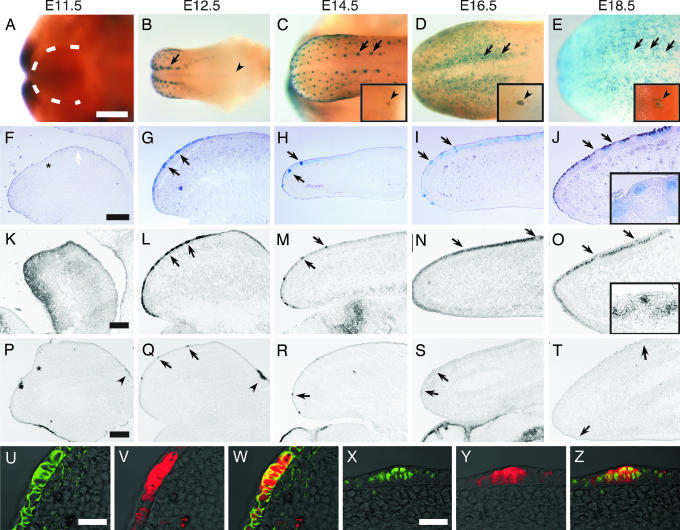
Fig. 1.
Wnt/β-catenin signaling elements are expressed in the developing tongue. Expression in the developing tongue of β-galactosidase (β-gal) (A–J, V, and Y), Lef1 (K–O and X), Wnt10b (P–T), and β-catenin (U) was detected by X-Gal staining in TOPGAL mice (A–E), by in situ hybridization (K–T), or by immunofluorescence (U–Z) in wild-type mice during developmental stages E11.5–E18.5. At E11.5, β-gal activity is absent from the lateral swelling (within dotted line in A and the white arrow in F). β-gal activity first appears in the placode of the dorsal tongue at E12.5 (B and G, arrows). Arrowheads in B and Insets in C–E indicate CV papillae. β-gal activity is absent from CV papillae until E14.5 (C). Lef1 expression is detected at E11.5 in both the mesenchyme and epithelium of the tongue (K), then increases in the fungiform papilla placode at E12.5 (L). At later stages, Lef1 expression continues in the fungiform papillae but at E16.5, and thereafter Lef1 also is highly expressed in the developing filiform papillae (N and O), whereas its expression still remains in fungiform papillae at later stages (O Inset) in accordance with β-gal expression (compare with J Inset). Wnt10b expression is first seen at E11.5–E12.5 in the developing CV papillae (P and Q, arrowheads), then at E12.5 in the developing placode (Q), and continues to be expressed through E14.5 (R), but its expression declines by E16.5 (S). Fluorescent micrographs of E12.5 tongue placode sections from TOPGAL mice show that β-gal expression (V and Y) associates with β-catenin (U) and Lef1 (X). W and Z are merged images of U and V and X and Y, respectively. Asterisks in F, K, and P indicate the border between the mandible and developing tongue. (Scale bars: A–E, 500 μm; F–T, 200 μm; U–Z, 100 μm.)

