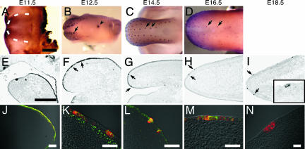
Fig. 2.
Shh and Wnt/β-catenin signaling elements are expressed similarly in the developing tongue. Expression in the developing tongue of Shh (A–I), Shh plus Lef1 (J–L), and Shh plus β-gal (M and N) was detected by whole-mount immunostaining (A–D), in situ hybridization (E–I), or double immunofluorescence (J–N) in wild-type (A–L) or TOPGAL (M and N) mice during developmental stages E11.5–E18.5. The dotted line in A indicates the border between the lateral swelling and the mandible. Arrows in B and F indicate the placode; arrows in C and D indicate fungiform papillae closest to the median sulcus. The arrowhead in B indicates the CV papilla. The Inset of I is a higher magnification view of a fungiform papilla. The pattern of expression of Shh is similar to that of β-gal during fungiform papillae formation (compare B–D with Fig. 1 B–D; also compare F–H with Fig. 1 G–I). The asterisk in E indicates the border between the mandible and developing tongue. Merged fluorescent micrographs of tongue placode sections from E12.0 (J), E12.5 (K and M), and E13.5 (L) stage mice show coexpression of Shh (red; J–N) with Lef1 (green; J–L), or β-gal (green; M). The CV papilla at E12.5 (N) expresses Shh (red) but not β-gal (green). The component single fluorescence images corresponding to merged images K–M are shown in supporting information (SI) Fig. 6. (Scale bars: A–I, 500 μm; J–N, 50 μm.)

