
Fig. 6.
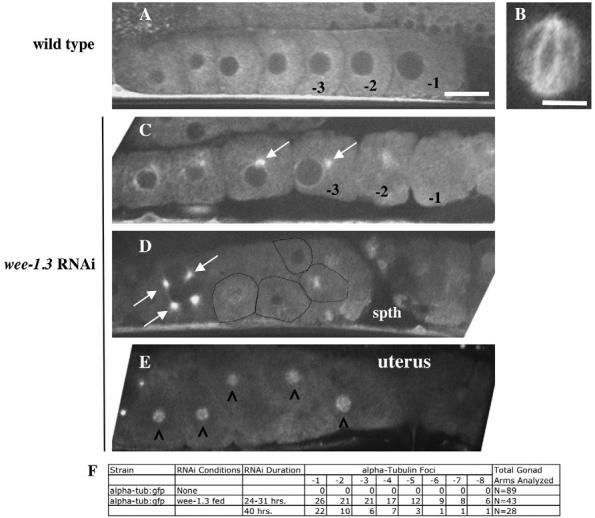
WEE-1.3 depletion results in the appearance of aberrant microtubules in developing oocytes. (A-E) Live α-tubulin::GFP animals were subjected to wee-1.3 RNAi. (A,B) Untreated animals. (A) Oocytes with typical cytoplasmic tubulin cytoskeletons. (B) A wild-type meiotic spindle; such a structure is normally observed in the oocyte upon fertilization in the spermatheca, or in the +1 embryo in the uterus, and is never observed in WEE-1.3-depleted oocytes in the spermatheca or uterus. (C-E) WEE-1.3-depleted oocytes in the oviduct (C,D) or the uterus (E). White arrows mark the perinuclear tubulin foci. Oocytes in D are outlined in black. (E) Tubulin clouds (marked by carets) form around the coalesced chromosomes of WEE-1.3-depleted oocytes that fail to be fertilized but nonetheless end up in the uterus. The spermatheca is to the right in each gonad shown. Scale bars: in A, 20 μm for A,C-E; 10 μm in B. (F) Quantitation of the number of oocytes with tubulin foci scored by oocyte position in the proximal gonad. spth, spermatheca.