
Figure 2.
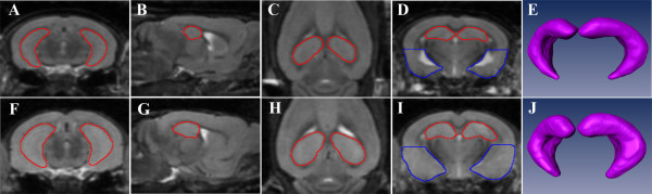
Segmentation of wild type and Kv1.1 null hippocampus and ventral cortex. The borders of the hippocampus (red) were drawn in three dimensions. Examples are shown for coronal (A, F), sagital (B, G) and horizontal (C, H) planes. The segmentation resulted in a 3D surface reconstruction of the hippocampus (E, J). The ventral cortex volume was derived from ventral cortex area (blue) measured in four coronal sections evenly distributed from 1.2 to 2.5 mm posterior to Bregma (D, I). Note the difference in size between wild type (top, i.e. A, B, C, D, E) and Kv1.1 null (bottom, i.e. F, G, H, I, J).