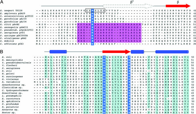
Fig. 5.
Putative arginine finger residues in ParG and MinE proteins. (A) N-terminal regions of ParG homologs. Blue, putative arginine finger residue; magenta, alanine/serine patch. Region 17–23 in TP228 ParG, which is less mobile than the remainder of the N-terminal region, is boxed. Secondary structure features in TP228 ParG are shown above (6, 7). (B) N-terminal regions of MinE homologs. Conserved arginines that are candidate arginine finger residues are outlined in blue. Secondary structure features in E. coli and Neisseria meningitidis MinE are shown (21, 22, 36). The E. coli MinE N-terminal domain (residues 1–35) is predicted to consist of an extended or nascent helix (22).

