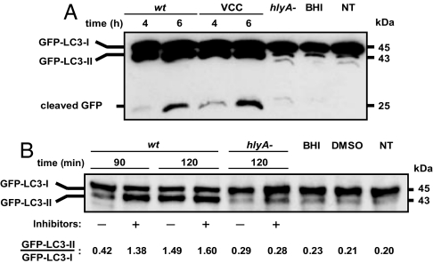
Fig. 2.
LC3 processing upon VCC intoxication. (A) VCC enhances LC3 processing. CHO-LC3 cells were incubated with filter-sterilized culture supernatants from Vc-wt or Vc-hlyA− (1:100 dilution), with purified VCC (60 pM), or with noninoculated BHI medium (1:100 dilution). After the indicated incubation times, protein cell extracts were subjected to SDS/PAGE and the GFP-LC3 forms (I and II) or free cleaved GFP were detected by Western blot analysis, as described in Materials and Methods. NT, nontreated. (B) Inhibition of lysosomal proteases leads to LC3-II accumulation during VCC-induced autophagy. Shown is a representative immunoblot of CHO-LC3 cells treated with sterile culture supernatants from Vc-wt or Vc-hlyA− (1:100 dilution), with or without previous incubation with pepstatine A (10 μg/ml) and E64d (10 μg/ml) 2 h before stimuli. As control, cells treated only with 1:100 dilution of BHI, 0.1% vol/vol DMSO (120 min each), and NT cells were assayed. Protein extracts were obtained and analyzed by Western blotting using an anti-GFP antibody.

