Fig. 1.
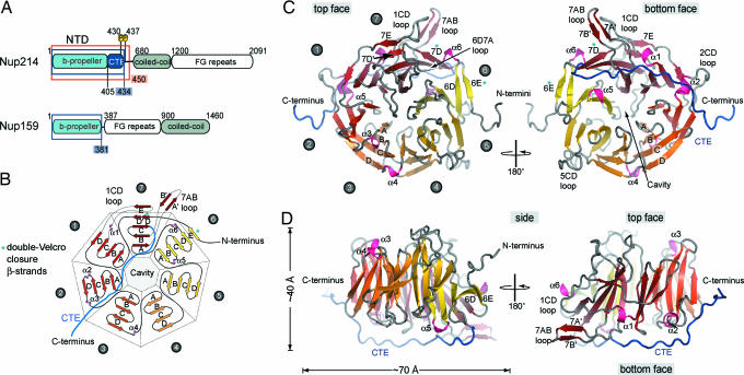
The structure of the NTD of human Nup214. (A) Domain structure of Nup214 and Nup159. The construct used for crystallization is boxed red, and two phosphorylation sites of the NTD are indicated. Residues observed in the crystal structures are boxed in blue. (B) Schematic representation of the NTD structure. The blades of the β-propeller are labeled from 1 to 7. The CTE is shown in blue, and β-strands forming the double-Velcro closure are indicated with an asterisk. (C) Ribbon representation of the NTD structure. A 180°-rotated view is shown on the right. As a reference, the strands of blade 3 are labeled A–D. The blades of the β-propeller and the CTE are labeled as in B. The helical insertions are shown in pink. (D) Ribbon representation of side views of the structure of the NTD. The view on the right is rotated by 180°.

