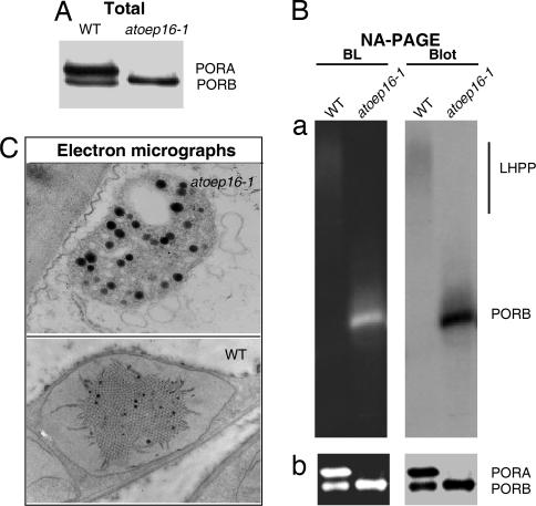
Fig. 6.
POR expression in etiolated Atoep16-1 and wild-type seedlings. (A) PORA and PORB protein levels in Atoep16-1 and wild-type etioplasts determined by SDS/PAGE and Western blotting. (B) Detection of light-dissociable PORA:PORB-Pchlide-NADPH supracomplexes indicative of LHPP in solubilized membrane fractions of Atoep16-1 and wild-type etioplasts before (a) and after (b) a 1-msec flash of white light. Protein detection was made by nondenaturing, analytical PAGE (NA-PAGE) and blue light (BL)-induced pigment autofluorescence (Left) and protein gel blot (Blot) analysis (Right) using a POR antiserum. (C) Transmission electron micrographs of Atoep16-1 and wild-type etioplasts. Black dots represent plastoglobules formed in excess in Atoep16-1 versus wild-type etioplasts.

