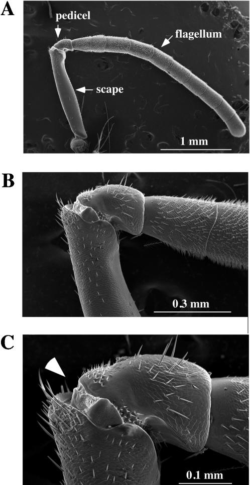
Figure 1.
External morphology of the honey bee antenna. The honey bee antenna was examined by scanning electron microscopy with different magnifications (A; x40, B; x150, C; x 300). Two proximal antennal segments (scape and pedicel) and the ten segments of the flagellum are indicated by arrows in A. Arrowhead in C indicates the position of electrode insertion for SEP recordings. The scale of each panel is shown by a white bar.

