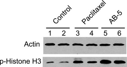
Fig. 3.
Assessment of phospho-histone H3 in xenografted tumors. Nude mice were injected at an s.c. site with 107 PC3 cells in the left flank. Eight days later, the mice were randomized into three groups of two mice each and treated with vehicle control (lanes 1 and 2), 20 mg/kg paclitaxel (lanes 3 and 4), and 20 mg/kg AB-5 (lanes 5 and 6). The mice were killed 12 h later, and extracts from the tumors were immunoblotted with antiphosphorylated histone H3 and antiactin antibodies.

