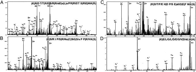
Fig. 2.
MS/MS spectra of four phosphorylated Lys-C peptides identified by ETD. Phosphopeptides with charge states of + 5 (A), +4 (B), +3 (C) and +2 (D) are shown. The four peptides originate from splicing factor, arginine/serine-rich 2 interacting protein, splicing coactivator subunit SRm300, Bcl2-associated transcription factor 1 and DnaJ homolog, subfamily C, member 9, respectively. The peptide sequence with the fragmentation pattern is shown in each panel. The signs: “/”, “ /”, and “|” designate that the C-terminal type fragments, N-terminal fragments, or both types of fragments, respectively, were identified. For all spectra, except A, the intensity axis has been enlarged by a factor of ≈5. Fragment ions resulting from charge stripping of the precursor ion are assigned with charge states (in bold). Small letters indicate phosphorylated residues.

