
Figure 3.
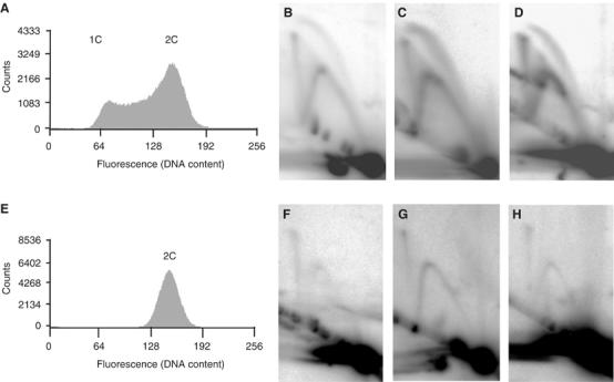
X-shaped molecules are present at all three replication origins and persist into the nonreplicating stationary phase. (A) Fluorescence histogram of an asynchronous replicating S. solfataricus culture (optical density (A600) of 0.3). The fluorescence values in the histogram feature two peaks corresponding to one and two copies (1C and 2C) of the genome, respectively. The histogram region between the two peaks includes those cells in DNA synthesis (S) phase. (B–D) 2D gel analysis of the three origins of replication using DNA isolated from the replicating population analysed in panel A (panel B depicts oriC1 (AccI digest), panel C illustrates oriC2 (BamHI/XcmI digest) and panel D demonstrates oriC3 (PvuII digest)). Bubble-, fork- and X-shaped molecules are present at each origin. (E) Fluorescence histogram of a stationary phase S. solfataricus culture. (F–H) 2D gel analysis of the three origin loci using DNA isolated from the stationary phase population (restriction fragments are identical to panels B–D). Although bubble-shaped molecules are no longer detectable, X- and Y-shaped species persist.