
Figure 3.
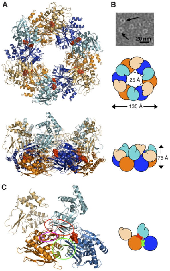
Hexameric ring structure, assembly, AMP-PNP binding, alternating subunit conformations, and subunit interactions. (A) AfGspE hexamer assembly and fold shown as ribbons and as schematic shapes viewed from top and side. Bound AMP-PNP (red, CPK spheres) in closed form molecules (blue, light blue NTD) and alternate open form molecules (orange, light orange NTD). (B) Electron micrograph of negatively stained AfGspE proteins with AMP-PNP. Ring structures are indicated by black arrows. (C) Subunit-subunit interactions with domain colors as in A, and encircled residues for N2:C1 (red), for C1:C1 (green), and N1:C2 (magenta) interactions.