
Figure 5.
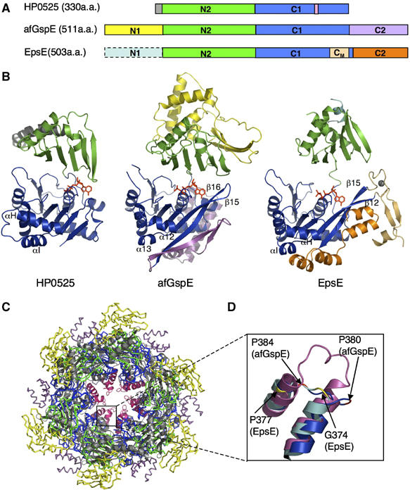
Secretion ATPase structural comparisons, subdomains, and a conserved functional core. (A) Schematic view of the three secretion ATPase structures. (B) Overall structures for HP0525 (left), afGspE (center), and EpsE (right) shown as ribbons with bound nucleotide (red sticks). Similar N2 (green) and C1 (blue) subdomains form the conserved core flanked by variable HP0525 N-terminal helix (gray), afGspE N1 (yellow) and C2 (magenta), and EpsE Zn2+ (gray sphere) region of the CM subdomain (light orange) and C2 (orange). (C) Structural comparison between afGspE (ribbons colored as in A) and HP0525 (gray) hexamers with pink HP0525 αH-I insertions. (D) Close up comparison of the closed site structure for HP0525 αH-I insertions (pink), afGspE α12–13 (blue), and EpsE (cyan). Colored residues kink the loop between helices in afGspE (red) and EpsE (yellow).