
Figure 4.
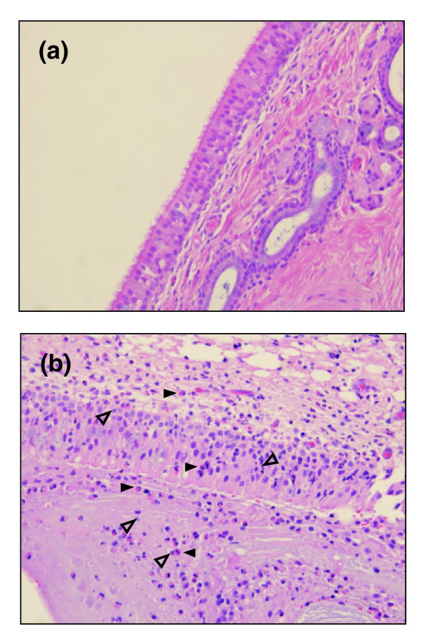
Representative sections of nasal mucosa fromewes 14 days post-inoculation with ovine or human B. parapertussis. (Hematoxylin and eosin stain, 400× magnification.) (a) Ewe inoculated with ovine strain. The epithelium has intact epithelial cells. The section lackssignificant infiltrates of inflammatory cells. (b) Ewe inoculated with human strain. The epithelium is covered by mucinous materialadmixed with neutrophils, eosinophils, necrotic cell debris, and seroproteinaceous fluid. Within the epithelium and lamina propria are moderate infiltrates of neutrophils (open arrowheads)and eosinophils (filled arrowheads). The lamina propria is moderately expanded by edema.