
Figure 2.
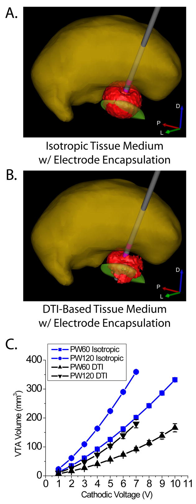
Volume of tissue activated by deep brain stimulation. VTA shape and size differed between isotropic (A) and DTI-based (B) tissue mediums, resulting in differential activation of surrounding anatomical structures. Both models included a tissue encapsulation layer around the electrode shaft, and volumes generated under the two conditions were matched for electrode impedance. C) Average VTA volume +/− standard deviation for all electrode contacts as a function of stimulus voltage for stimulus pulse widths of 60 μsec and 120 μsec.