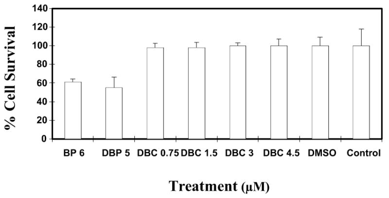
Fig. 3.

Cytotoxicity assay using MCF-7 cells in culture. MCF-7 cells were exposed to varying doses (0.75-4.5 μM) of DBC. Cells were also treated with BP and DBP at known cytotoxic doses of 6 and 5 μM, respectively, for comparison. Each data point represents the mean of four wells (mean ± SD). The percent cell survival is given in comparison to untreated cells.
