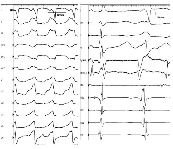
Figure 1.

Left panel: Surface 12-lead electrocardiogram of ventricular tachycardia (cycle length= 485 ms) observed during the ablation procedure (that was identical to the clinical tachycardia). Right panel: Intracardiac tracing of PVC’s mapped during ablation. HRAd: right atrial catheter, His px; His bundle proximal, His md: His bundle mid, His ds: His bundle distal, Rva: RV apex, Epi Abld: distal pole of epicardial ablation catheter, Epi Abl p: proximal pole of the epicardial ablation catheter
