Figure 2.

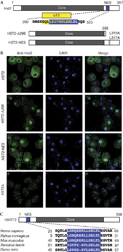
Hst2 localization is controlled by a leucine-rich nuclear export sequence. (A) A nuclear export sequence (NES; blue) was identified using the NetNES 1.1 prediction algorithms. The Hst2 α13 helix is shown in yellow. (B) Mutations in the NES region caused Hst2 to accumulate in the nucleus. Hst2 localized primarily to the cytoplasm; HST2-Δ298 and HST2-NES localized primarily to the nucleus. The hst2Δ cells illustrate low background staining. All strains are hst2Δ, bearing 2μ constructs. Immunofluorescence is as in Fig 1. DAPI, 4′,6-diamidino-2-phenylindole. (C) Alignment of NESs from vertebrate SIRT2 (hSIRT2) proteins. Blue boxes indicate NESs identified using NetNES 1.1. Multiple sequence alignments were created in Clustal W.
