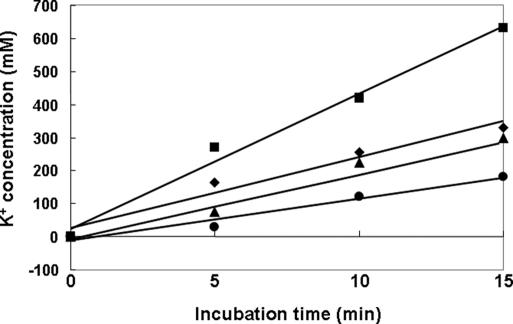
Fig. 5.
Analysis of intracellular accumulation of K+. Strains MG1655, CR201, and CR301 grown in K0 medium were harvested, washed, and resuspended in K20 minimal medium containing 0.5% glucose. Cells were harvested by filtration at the indicated time points and processed for analysis of K+ content by employing inductively coupled plasma/optical emission spectrometry (ICP-OES) as described in Materials and Methods. MG1655, diamonds; CR201 (ptsO), triangles; CR301 (ptsN), squares; and CR301/pCR3(H73A), circles.

