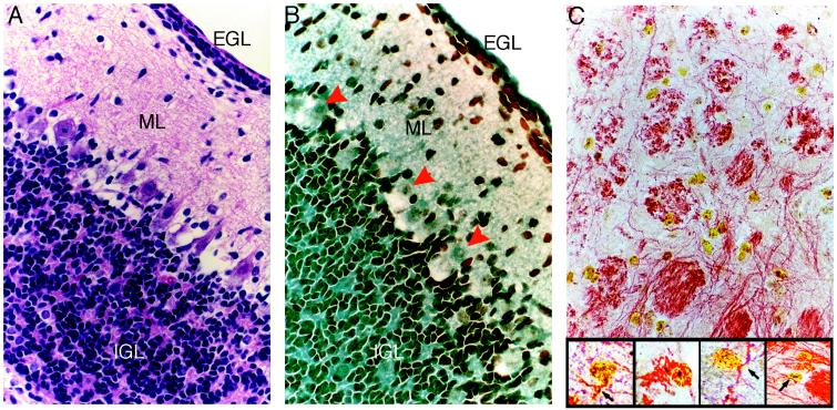
Figure 6.
Localization of BrdUrd-labeled MSCs in cerebellum. Hematoxylin/eosin (A) or anti-BrdUrd (B) staining of serial sections reveal MSCs within the EGL, molecular layer (ML), and IGL of the cerebellum. Red arrowheads indicate negative staining of Purkinje cells. (C) MSCs in the reticular formation of the brain stem triple-labeled with anti-BrdUrd, anti-GFAP, and anti-neurofilament. (×400.) (Inset) Higher magnification reveals neurofilament staining (red reaction product) in the cytoplasmic processes of numerous BrdUrd (yellow reaction product)-labeled MSCs. (×1,000.)

