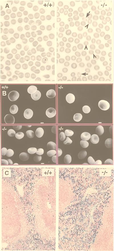
Figure 5.
(A) Bright field light microscopy. Arrows indicate rounded elliptocytes; arrowheads indicate microspherocytes. (B) Scanning electron microscopy demonstrates that −/− RBCs are smaller and spherocytic compared with +/+ RBCs. (Bar = 1 μm.) (C) Increased iron deposition (dark blue color) in spleen from −/− mice compared with +/+ mice.

