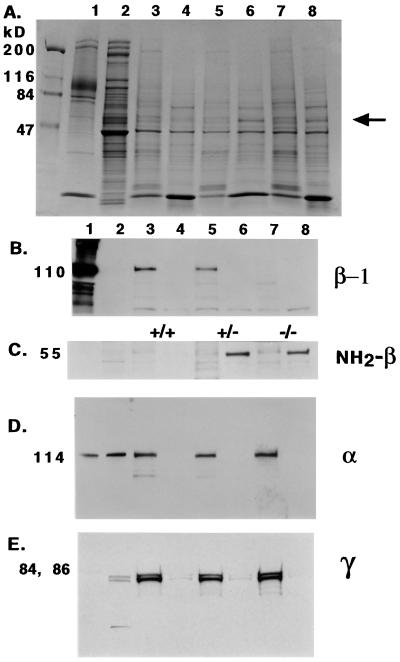
Figure 8.
Western blotting of spleen homogenates. Lanes: 1, human RBC ghosts; 2, human platelets; 3–8, spleen homogenates, separated into 30,000 × g pellet (P) and supernatant (S) fractions: 3, +/+ P; 4, +/+ S; 5, +/− P; 6, +/− S; 7, −/− P; 8, −/− S. Gel in A is Coomassie blue-stained to demonstrate protein loading. Gels in B–E are stained with the antibodies indicated on the right of the panels. The arrow marks the 55-kDa band in lanes 6 and 8 that is immunoreactive to NH2-β antibody. β-1 is not detected in −/− samples, but α and γ are detected in all pellet samples.

