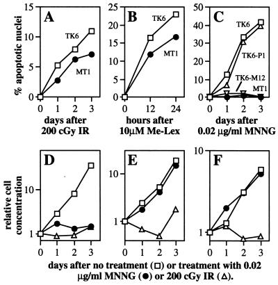
Figure 4.
Apoptosis and cell growth after treatment of human lymphoblastoid cells. Apoptosis was quantitated by nuclear morphology; for A–C, TK6 (□), MT1 (●), TK6 containing control vector, TK6-P1 (▵), TK6 expressing MGMT, TK6-M12 (▿). Me-Lex, methyl-lexitropsin. Cell density was monitored for TK6-P1 (D), TK6-M12 (E), and MT1 (F) after exposure to MNNG and IR.

