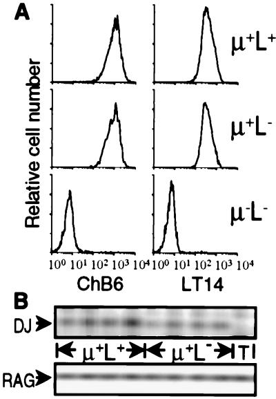
Figure 2.
μ+L− cells in the bursa of RCAS-Tμ-infected neonatal chicks are B lineage cells. (A) Bursal cells from neonatal chicks infected as day 3 embryos with RCAS-Tμ were stained for the expression of μ, IgL, and ChB6 or LT14. Histograms of 10,000 cells gated on μ and L expression and stained for ChB6 or LT14 are shown. (B) Bursal cells from neonatal chicks infected as day 3 embryos with RCAS-Tμ were stained and FACS-sorted based on the expression of μ and L. Typical purity of the sorted populations was >98%. Genomic DNA was PCR-amplified from 1,500 sorted μ+L+ and μ+L− bursal cells from four individual RCAS-Tμ-infected chicks and from unstained thymocytes (T) for the presence of DJH rearrangements and RAG2.

