Figure 2.
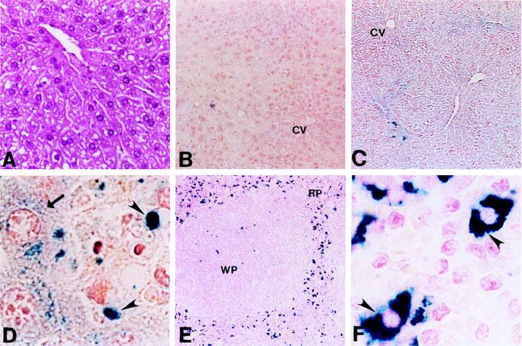
(A) Hematoxylin/eosin stain of liver from a representative 1-year-old Cp−/− animal (×17). (B) Perls’ stain of liver section from a representative 1-year-old Cp+/+ mouse (×8.5). CV, central vein. (C) Perls’ stain of liver section from Cp−/− littermate (×8.5). (D) High-power view from C (×100). Arrow indicates iron accumulation in hepatocyte; arrowheads indicate Kupffer cells. (E) Perls’ stain of Cp−/− spleen from 1-year-old mouse (×8.5). RP, red pulp; WP, white pulp. (F) High-power view from E (×100). Arrowheads indicate iron within splenic reticuloendothelial cells.

