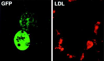
Figure 4.
Hepatocyte-specific transduction by recombinant hepadnaviruses. (Left) Infection of PDH cultures with rDHBV-GFP at an moi of 100 vp per cell for 24 hr. Transduced hepatocytes were detected by GFP expression at day 6 p.i. (Right) Sinusoidal endothelial cells surrounding the same hepatocytes were identified by receptor mediated uptake of red-fluorescent acetylated low density lipoprotein (LDL). Hepatocyte specificity is shown by the absence of GFP expression in the sinusoidal endothelial cells. (Confocal microscopy, ×63 lens.)

