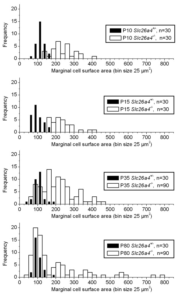
Figure 3.
Reorganization of marginal cells. Apical surface areas of marginal cells were measured using images of whole-mounts of stria vascularis as shown in Fig. 2. Frequency histograms (bin size 25 μm2) of surface area measurements were constructed. The number (n) of cells contributing to each histogram is given. Note that surface areas in Slc26a4+/- mice of all ages were normally distributed and narrowly centered at ~ 100 μm2. The distribution of surface areas in Slc26a4-/- mice progressed during development from a distribution centered at ~ 240 μm2 to a wide multimodal distribution with peaks centered at ~ 100 and ~ 240 μm2.

