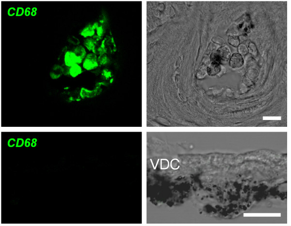
Figure 7.

Controls for immunohistochemistry. Controls for CD68 immunohistochemistry were obtained from cryosections of the cochlea. Top: bone marrow cells congregated in cavities of the bony cochlear wall served as a positive control. Bottom: heavily pigmented cells in the connective tissue underneath vestibular dark cells (VDC) served as a negative control. Left: confocal immunohistochemistry of CD68. Right: corresponding bright field images. Scale bars represent 10 μm.
