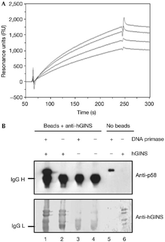
Figure 2.

Physical interaction between human GINS and human DNA primase. (A) An overlaid plot of sensorgrams was obtained by fluxing the human heterodimeric DNA primase at various concentrations over an hGINS-immobilized sensor chip, as described in the Methods (lower to upper curve: the DNA primase was at 25, 50, 100 and 200 nM, respectively). (B) Immunoprecipitation experiments were carried out using protein A Sepharose beads conjugated with anti-hGINS antibodies and the following samples: a mixture of purified recombinant hGINS and the heterodimeric DNA primase (lane 1), hGINS alone (lane 2) and human DNA primase alone (lane 3), as described in the Methods. A sample of beads conjugated with antibodies (lane 4), human DNA primase (100 ng, lane 5) and hGINS (200 ng, lane 6) were run as control. Western blot analysis was carried out by using anti-human primase p58 and anti-hGINS antibodies, as described in the Methods. hGINS, human GINS; RU, resonance units.
