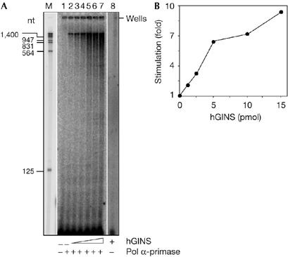
Figure 4.

Human GINS stimulates DNA synthesis by polymerase α-primase on singly primed M13 mp18 single-stranded DNA. (A) Reaction mixtures (volume 10 μl) contained singly primed M13mp18 single-stranded DNA (25 fmol), 18 μM dATP, 18 μM dGTP, 18 μM dTTP, 6 μM dCTP and [α-32P]dCTP (1.25 μCi) and, where indicated, polymerase (pol) α-primase (70 fmol). Lane 1, no enzyme control; lane 2, pol α-primase alone; lanes 3–7, pol α-primase in the presence of 1.27, 2.55, 5.1, 10.2 and 15.3 pmol of hGINS, respectively. Samples were incubated at 37°C for 15 min. Reaction products were run on a 6% polyacrylamide sequencing gel, which was analysed using a phosphorimager apparatus. Lane M contains EcoRI–HindIII-digested λ DNA as the size marker (the exposure of this lane was shorter for identifying the position of the marker). Lane 8 contains a control reaction carried out with 1.5 μg of hGINS without pol α-primase and run on a different gel. (B) Reaction products longer than 125 nucleotides detected in lanes 2–8 of the gel shown in (A) were quantified by using a phosphorimager, as described in the Methods. The intensity of the signal in each lane was normalized to the value calculated for the reaction carried out in the absence of hGINS. hGINS, human GINS.
