
Figure 1.
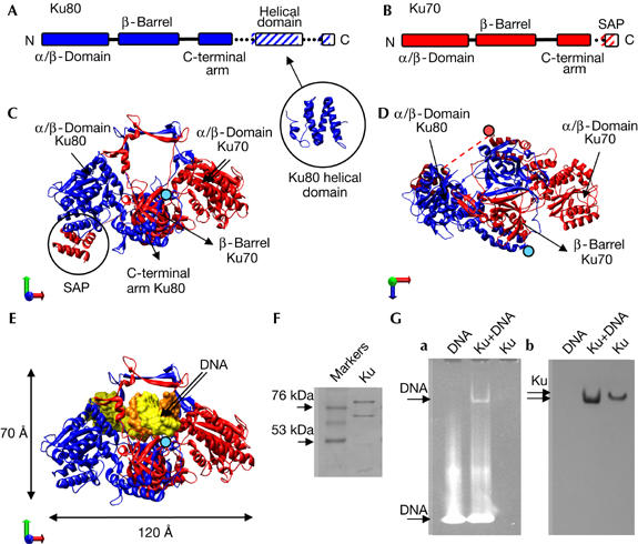
Purification of the Ku70–Ku80 dimer. (A) Schematic representation of the domain organization of Ku80. Ku80-CTD contains a helical domain (PDB 1Q2Z; Harris et al, 2004). (B) Schematic representation of Ku70. (C,D) Two views of the Ku70–Ku80 atomic structure (PDB 1JEQ; Walker et al, 2001). Ku80 and Ku70 subunits are coloured blue and red, respectively. In (D), a dotted line represents the connection between the core of Ku70 and the SAP domain, not solved in the structure. (E) Front view of the Ku-DNA atomic structure (PDB 1JEY). In (C–E), the last amino acid of each carboxy-terminal arm, from which the subunit-specific C-terminal domains emerge, is indicated with either a blue (Ku80) or a red (Ku70) filled circle. (F) SDS gel of purified Ku70–Ku80. (G) DNA interaction assay. (a) 6% native polyacrylamide gel electrophoresis gel developed with UV to show the presence of DNA. (b) Western blot of the native gel using a monoclonal antibody against Ku70. SDS, sodium dodecyl sulphate.