
Figure 1.
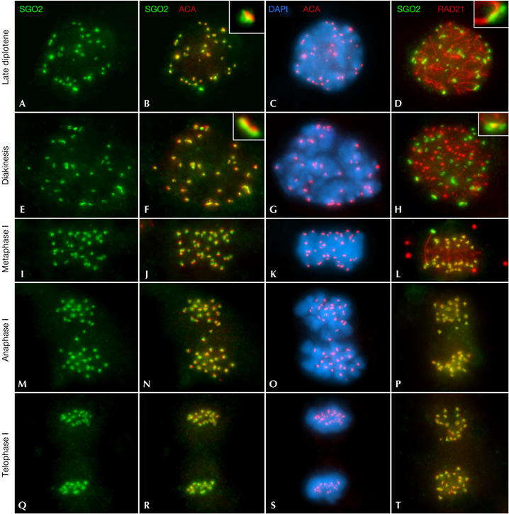
SGO2 distribution during meiosis I. Double immunolabellings of SGO2 (green) with kinetochores (ACA; red; columns 1–3); or RAD21 (red; column 4); and counterstaining with 4,6-diamidino-2-phenylindole (DAPI; blue; column 3). (A–D) Late diplotene; (E–H) diakinesis; (I–L) metaphase I; (M–P) anaphase I; and (Q–T) telophase I spermatocytes. All images are projections of different focal planes throughout the cell volume. ACA, anti-centromere autoantibody.