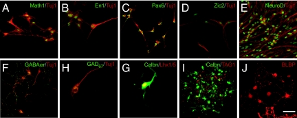
Fig. 2.
Differentiation of ES cells into cerebellar granule cells. After 16 DIV, ES cells were cultured in N2 Supplement-B and B27 (StemCell Technologies) as well as FGF8b, WNT1, WNT3a, BMP7, GDF7, BMP6, SHH, BDNF, and NT3. The latter factors induced expression of markers of dorsal neurons (green) MATH1 (A), the cerebellar territory EN1 (B), and granule neurons PAX6 (C), ZIC2 (D), NEUROD (E), and GABAα6r (F) as well as markers for Purkinje cell GAD67 (G), LHX1/5 (red, H), and CALB1 (green, H and I) and Bergman glia BLBP (red, J). The postmitotic neuronal markers (red) TUJ1 (A–F) and TAG1 (I) are also shown. (Scale bar: 20 μm for A–H and J and 40 μm for I.)

